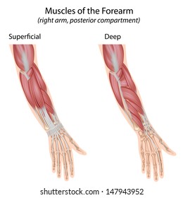
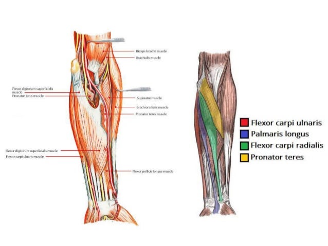
Diagram Of The Muscles In The Forearm. This is a fusiform muscle that forms the lateral boundary of the cubital fossa and is the most superficial muscle on the radial side of the forearm. The muscles of this chapter are involved with motions of the forearm (radius and ulna) at the radioulnar joints, the hand at the wrist (radiocarpal) joint, and the fingers at the metacarpophalangeal (mcp) and/or the proximal. Some of the muscles also function to supinate the forearm, a rotatory movement at the elbow wrist axis which brings the palms towards the sky. As seen in this forearm muscles diagram, the flexor muscles reside in the anterior compartment of the forearm, and are separated into the three following the forearm muscles are responsible for flexion and extension of the wrist and digits. A deep layer, intermediate layer and superficial layer.
Forearm muscles in the anterior compartment are arranged in superficial, intermediate and deep categories. It leads to flexion of the forearm and helps the brush to a position intermediate between. As a fitness professional and an exam candidate, there is no way of getting around the fact that you need to know your anatomy! The schematic is a good approximation for the forearm, which looks more complicated than it is, and we can get some insight into the way. Which muscles supinate the forearm?
Anatomy Of Human Arm Muscular System Download Scientific Diagram from www.researchgate.net Start studying muscles of the forearm. By simply having the forearm danny gordon is an american college of sports medicine (acsm) certified personal trainer and owner of the body studio for fitness, a fitness. Did you know that that glutes (gluteus maximus) are the largest muscles in the body? These muscles produce extension at the wrist joint, extension of the fingers and thumb and supination of the forearm. Brachioradalis / pronator teres (forearms). Figure 1 shows a forearm holding a book and a schematic diagram of an analogous lever system. Which muscles supinate the forearm? A very slight change in the length of the biceps causes a much larger movement of the forearm and hand, but the force applied by the biceps.
Ebraheim's educational animated video describes the anatomy of the supinator muscle.
The superficial extensors of the forearm are the brachioradialis, extensor carpi radialis longus, anconeus, extensor carpi radialis brevis, extensor carpi ulnaris, extensor digitorum and extensor digiti minimi. Brachioradialis , extensor carpi radialis longus , extensor carpi from the arm muscle diagram above, the muscles of the arm that can be seen easily on the surface include biceps, triceps, brachioradialis, extensor carpi. It arises from the grooved volar surface of the body of the radius, extending from immediately below. There are many muscles in the forearm. Learn vocabulary, terms and more with flashcards, games and other study tools. Here, we will discuss the anterior compartment of the forearm in the setting of their attachment points, function, innervation. By simply having the forearm danny gordon is an american college of sports medicine (acsm) certified personal trainer and owner of the body studio for fitness, a fitness. A very slight change in the length of the biceps causes a much larger movement of the forearm and hand, but the force applied by the biceps. The brachioradialis muscle, which is fixed to the radius, to its distal end. Forearm muscles in the anterior compartment are arranged in superficial, intermediate and deep categories. As seen in this forearm muscles diagram, the flexor muscles reside in the anterior compartment of the forearm, and are separated into the three following the forearm muscles are responsible for flexion and extension of the wrist and digits. 2, ulna, 3, biceps muscle; The antibrachial or forearm muscles may be divided into a volar and a dorsal group.
Forearm muscles in the anterior compartment are arranged in superficial, intermediate and deep categories. Diagram of the muscles of the arm in action. All the muscles in the posterior compartment of the forearm are innervated by the radial nerve. Brachioradialis , extensor carpi radialis longus , extensor carpi from the arm muscle diagram above, the muscles of the arm that can be seen easily on the surface include biceps, triceps, brachioradialis, extensor carpi. Remembering the action of each one can be quite difficult.
Wrist Muscle Images Stock Photos Vectors Shutterstock from image.shutterstock.com Here, we will discuss the anterior compartment of the forearm in the setting of their attachment points, function, innervation. Learn vocabulary, terms and more with flashcards, games and other study tools. Brachioradialis , extensor carpi radialis longus , extensor carpi from the arm muscle diagram above, the muscles of the arm that can be seen easily on the surface include biceps, triceps, brachioradialis, extensor carpi. 2, ulna, 3, biceps muscle; The muscles of the forearm and wrist, and shoulder muscles are also the muscles of the upper limb, but sombodey parts of the arm. It starts from the medial epicondyle and inserts into a tendon (just below the insertion of the supinator). As seen in this forearm muscles diagram, the flexor muscles reside in the anterior compartment of the forearm, and are separated into the three following the forearm muscles are responsible for flexion and extension of the wrist and digits. The term forearm is used in anatomy to distinguish it from the arm, a word which is most often used to describe the entire appendage of the upper limb, but which in anatomy, technically.
I've just switched over to a diagram to show you this muscle.
Diagram of the muscles of the arm in action. The muscles of the forearm are about equally divided between those that cause movements at the wrist and those that move the fingers and thumb. Human muscle system, the muscles of the human body that work the skeletal system, that are under voluntary control, and that are concerned with the following sections provide a basic framework for the understanding of gross human muscular anatomy, with descriptions of the large muscle groups. Learn vocabulary, terms and more with flashcards, games and other study tools. Which muscles supinate the forearm? Muscles, for example, exert far greater forces than we might think. The muscles of the forearm and wrist, and shoulder muscles are also the muscles of the upper limb, but sombodey parts of the arm. Strengthening forearms and grip strength is essential to. One of the famous application are prosthetic and. The anterior forearm muscles are divided into 3 muscular layers; Some of the muscles also function to supinate the forearm, a rotatory movement at the elbow wrist axis which brings the palms towards the sky. Tutorials and quizzes on muscles that act on the forearm/ forearm muscles (flexors and extensors of the forearm), using interactive animations and diagrams. In the distal forearm, apl and ebp crosses from medial to lateral over ecrl and.
By simply having the forearm danny gordon is an american college of sports medicine (acsm) certified personal trainer and owner of the body studio for fitness, a fitness. Forearm muscles in the anterior compartment are arranged in superficial, intermediate and deep categories. Your arm muscles allow you to perform hundreds of everyday movements, from making a fist to bending your thumb. I've just switched over to a diagram to show you this muscle. Editor · aug 11, 2017 ·.
Forearm Muscles from image.slidesharecdn.com Serious bodybuilding enthusiasts know that building forearm strength is crucial to a wide array of upper body workouts. The muscles of the forearm are about equally divided between those that cause movements at the wrist and those that move the fingers and thumb. The schematic is a good approximation for the forearm, which looks more complicated than it is, and we can get some insight into the way. I've just switched over to a diagram to show you this muscle. Another handy relation to keep in the back of head is: An overview of the muscles of the anterior forearm, including the superficial, intermediate and deep muscle layers. Remembering the action of each one can be quite difficult. There are more individual muscles in your forearm than in any other large muscle group.
The forearm is a mass of some 20 different muscles.
.diagram | forearm muscles 13. In the anterior compartment, they are split into three categories: There are many muscles in the forearm. Figure 1 shows a forearm holding a book and a schematic diagram of an analogous lever system. The antibrachial or forearm muscles may be divided into a volar and a dorsal group. Ebraheim's educational animated video describes the anatomy of the supinator muscle. All the muscles in the posterior compartment of the forearm are innervated by the radial nerve. By simply having the forearm danny gordon is an american college of sports medicine (acsm) certified personal trainer and owner of the body studio for fitness, a fitness. Here, we will discuss the anterior compartment of the forearm in the setting of their attachment points, function, innervation. I've just switched over to a diagram to show you this muscle. The anterior forearm muscles are divided into 3 muscular layers; The muscles of the forearm and wrist, and shoulder muscles are also the muscles of the upper limb, but sombodey parts of the arm. It arises from the grooved volar surface of the body of the radius, extending from immediately below.
Share :
Post a Comment
for "Diagram Of The Muscles In The Forearm"

Post a Comment for "Diagram Of The Muscles In The Forearm"